- Phone
-
Address
Building 1, International Entrepreneurship Park, No.2 Shangdi Information Road, Haidian District, Beijing
Product Categories
- MetaMorph research grade imaging software
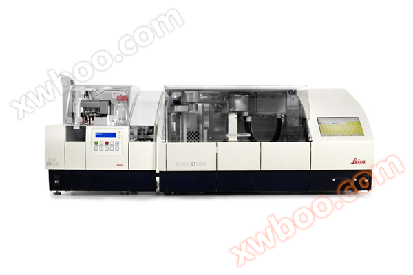
- Dyeing Machine
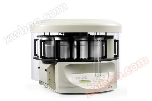
- Modular organization embedding system
- Slide scanner/digital pathology scanner
- Multi functional enzyme-linked immunosorbent assay (ELISA) reader
- Slicer/Oven
- Single function enzyme-linked immunosorbent assay (ELISA) reader
- Upright microscope series
- metallurgical microscope
- Digital Interactive Classroom
- Microscopic digital analysis system
- Number machine
- High connotation imaging analysis system
- Polarizing microscope
- Dehydration machine
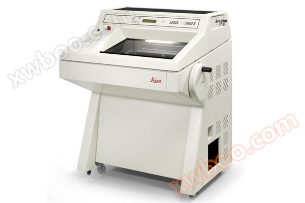
- Slicing machine
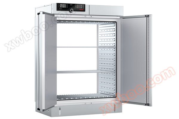
- Memmert oven
Beijing Yuechangxing Technology Co., Ltd
Ion imaging system
NegotiableUpdate on 04/01
- Model
- Nature of the Manufacturer
- Producers
- Product Category
- Place of Origin
Overview
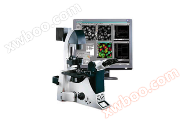
The MD ion imaging system is designed specifically for the proportional method and is equipped with powerful hardware control and image analysis software MetaFlour. It can be used in conjunction with patch clamp to monitor changes in cellular ions.
Product Details
Calcium ions are one of the most important ions in life activities. By measuring the concentration of free calcium ions in cells, researchers can obtain important information about life activities such as muscle contraction, nerve signal transduction, intercellular communication, and hormone responses. At present, the mainstream measurement methods for intracellular calcium ions in the world are electrical measurement and optical measurement. The electrical measurement method uses cell membrane clamp technology to determine the changes in cell membrane potential caused by calcium current. The light measurement method uses fluorescent probes that can specifically bind to calcium ions to measure changes in cell fluorescence intensity. If a standard solution is used to create a standard curve, the absolute concentration of intracellular calcium ions can even be measured, making it one of the most authoritative methods for measuring intracellular free calcium ion concentration in the world. The MD ion imaging system is designed specifically for the proportional method and is equipped with powerful hardware control and image analysis software MetaFlour. It can be used in conjunction with patch clamp to monitor changes in cellular ions.

main features
Long life xenon lamp light source, high stability, high brightness, can be used for 10000 hours.
●Color filter: Optional rotary switch, widely applicable, fast speed of 40-60 ms, versatile, capable of ion imaging and common fluorescence imaging.
●High speed switching, capable of meeting fast response experiments, with switching speed in milliseconds.
●MetaFluor/MetaMorph fluorescence ion concentration measurement and image analysis software: for measuring intracellular ion concentration at single or dual wavelengths, suitable for commonly used Ratio indicators such as Fura-2, BCECF, INDO-1, etc. It can simultaneously display raw wavelength images (up to four wavelengths), Ratio images (can simultaneously produce Ratio images for two different indicators), and curves of changes in optical density, Ratio, and ion concentration of different cells. The software can also control the shutter of the excitation light source to minimize the damage caused by prolonged exposure of the excitation light to cells. Image acquisition: Obtain up to five wavelengths of images per cycle; Background reduction function (each wavelength has an independent background); Shadow correction function, with independent shadow reference images for each wavelength.
●Run JOURNAL at specific time points during the experimental cycle, automatically run JOURNAL, TIME LAPSE 。 Calibration: Use the standard GRYNKIEWICZ equation for calibration; Calibrate using the selected curve fitting mode; Generate calibration images and directly display pH values, calcium ion concentrations, or other ion concentrations.
●Analysis: Obtain two different indicators, Ratio, for each image cycle; Log data into text files, Microsoft Excel, or other applications; Analyze multiple regions of interest, measure the intensity, strength, and HREHOLD area of each region of interest at various time points RATIOS、 Calibrated ion Concentration, etc.
application area
The ion imaging system of Molecular Devices includes high-precision and high-speed components that can perform high-speed ion imaging analysis and high-precision proportional imaging. Combined with the highest level MetaFluor software system, it can be used for fluorescence ratio measurement of various ions (Ca2+, Mg2+, Zn2+, K+, Cl -, Na+, pH) such as nerve cells, myocardial cells, skeletal muscle cells, pancreatic island cells, as well as single wavelength fluorescence measurement and fast FRET measurement.
Experimental examples
Fura-2 fluorescent dye is used to detect changes in calcium ions within nerve cells, muscle cells, or secretory cells.
Fura-2 is a ratio type indicator that undergoes spectral changes in both calcium free and calcium bound states. For example, in the calcium bound state, its excitation peak changes from 363 nm to 335 nm, while the emission peak shows no significant change. Therefore, it is usually excited twice with dual wavelengths (340 nm and 380 nm), and the ratio of the emission intensity obtained after excitation is the relative value of the calcium signal. Because the data results are presented in a ratio manner, they are not limited by experimental equipment, indicator loading concentration, cell type, or individual experimenter operation. At the same time, they can eliminate the influence of factors such as cell thickness changes and indicator redistribution changes inside the cell, truly reflecting the changes in intracellular calcium levels.