- Phone
-
Address
Building 1, International Entrepreneurship Park, No.2 Shangdi Information Road, Haidian District, Beijing
Product Categories
- MetaMorph research grade imaging software
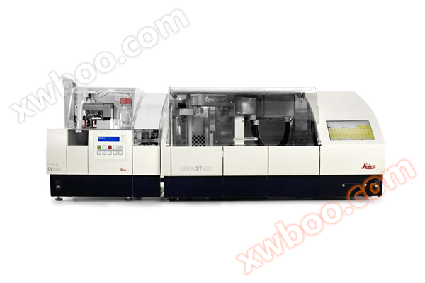
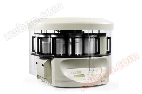
- Dyeing Machine
- Modular organization embedding system
- Slide scanner/digital pathology scanner
- Multi functional enzyme-linked immunosorbent assay (ELISA) reader
- Slicer/Oven
- Single function enzyme-linked immunosorbent assay (ELISA) reader
- Upright microscope series
- metallurgical microscope
- Digital Interactive Classroom
- Microscopic digital analysis system
- Number machine
- High connotation imaging analysis system
- Polarizing microscope
- Dehydration machine
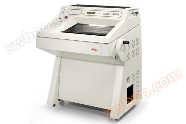
- Slicing machine
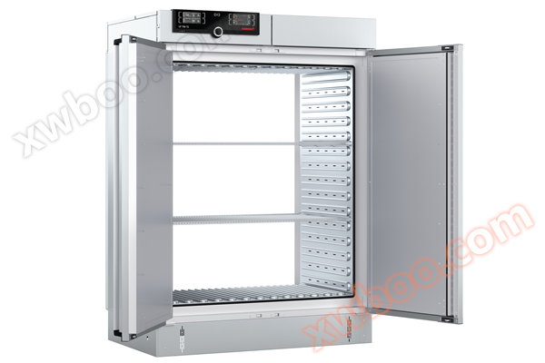
- Memmert oven
Beijing Yuechangxing Technology Co., Ltd

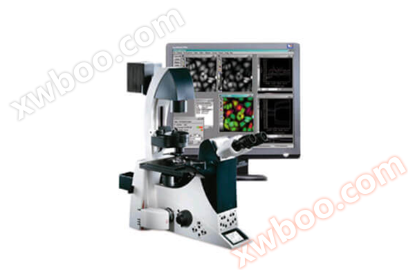
ImageXpress Micro Confocal Confocal High Content Imaging System
NegotiableUpdate on 04/01
- Model
- Nature of the Manufacturer
- Producers
- Product Category
- Place of Origin
Overview
The ImageXpress confocal high content imaging system combines acquisition speed, sensitivity, and operational flexibility to expand your research scope. Capture high-quality images of the entire biological organism, thick tissue, 3D cell sphere analysis, and cellular or intracellular events.
Product Details
ImageXpress Micro Confocal Confocal High Content Imaging Analysis System
The ImageXpress Micro Confocal confocal high content imaging analysis system is currently an extremely flexible high content system. Thanks to the clear characteristics of confocal imaging, experimenters can perform more accurate quantitative analysis on live cells or fixed cells. Using this system, experimenters can easily observe complex 3D models, such as 3D cell spheres, tissues, and even small model organisms as a whole, in order to further analyze more physiological related issues. This system can automatically and quickly obtain high-quality images of publication level from samples of 1-1536 microplates or tissue sections.

main features
Capable of automatically imaging 6-1536 porous plates, including Transwell plates, that meet or do not meet SBS standards for various specimens, specifications, and any bottom wall; And it can preview user made samples such as glass slides, cell chips, and tissue arrays through low magnification mirrors, customize sample specifications, and perform high-throughput automatic imaging and data analysis through high magnification mirrors.
●AgileOptixTM rotary confocal technology is an optical system patented by MD company, which can easily switch and set shooting modes to achieve optimal shooting and analysis results.
●Scientific grade sCMOS is currently the most advanced detector in microscopy imaging detection systems. SCMOS overcomes the disadvantages of low output rate and high power consumption of CCD, as well as the disadvantages of high noise and dark current of CMOS. It is a scientific grade image detector that integrates high resolution, high frame rate, high quantum efficiency, high sensitivity, high dynamic range, and wide field of view. (Combined with solid-state light engine light source to achieve the best imaging effect).
●Adopting magnetic levitation technology, it has fast speed, high accuracy, no wear, and no thermal fatigue.
●Laser hardware autofocus and software autofocus can be combined and used separately or simultaneously. Flexible application, suitable for various objective lenses and sample plates.
●By using a dedicated phase contrast objective lens, cells without fluorescent labeling can be observed and analyzed. Its phase contrast imaging module can obtain higher contrast unlabeled images, and the light source component has corresponding focusing lenses and phase contrast ring plates to support unlabeled phase contrast imaging, providing high contrast images and enabling cell recognition and tracking in unlabeled conditions.
●PowerCoreTM data parallel analysis function can call local and network idle CPU thread resources, which can improve image analysis speed by several times
●Various experimental parameters in complex 3D structures can be analyzed through simple mouse operations. Combined with MetaXpress's automatic Z-axis layer scanning, 3D display, and patented Best focus algorithm, porous plates can be used to achieve high-throughput 3D shooting and analysis in a streamlined manner.
●Immersion lenses: 20x, 40x, and 60x water mirrors improve the geometric accuracy during imaging and reduce the refractive index of high brightness fluorescence signals under lower exposure time conditions.
●High intensity laser light source: 5- or 8-high-intensity laser light source, expanding experimental adaptability.
●Confocal disc suitable for deep tissue penetration: The confocal disc module module that facilitates deep tissue penetration reduces crosstalk, enhances the suppression of defocused light, and strengthens the penetration of thick tissue.

application area
It can be used from cells, tissue sections to small model animals. The ImageXpress Micro Confocal system obtains the necessary images and quantitative analysis results for the experiment. Suitable for the vast majority of cellular life
Physical experiments quantitatively analyze various parameters of cells, transforming traditional imaging experiments from qualitative analysis to quantitative analysis. Suitable for research directions such as cell biology, toxicology, pharmacology, physiology, genetics and embryology, neurobiology, oncology, etc.

Experimental examples
Observation and analysis of 3D models, such as 3D cell spheres, tissues, and even small model organisms as a whole
Complex 3D cultured cell models are increasingly being used in drug development and basic research, mainly because compared to single-layer and 2D cultured models, 3D models are closer to the in vivo environment and can obtain more predictive and physiologically meaningful results. The IXM-C system provides a simple method for detecting 3D models, and even samples grown in thick Matrigel can obtain more accurate results. This imaging system can image various types of samples, including various specifications of Hanging Drop plates, U-shaped circular bottom well plates, and flat bottomed microporous plates. If equipped with environmental control components, it can dynamically track and detect cell health for a long time.