- Phone
-
Address
Building 1, International Entrepreneurship Park, No.2 Shangdi Information Road, Haidian District, Beijing
Product Categories
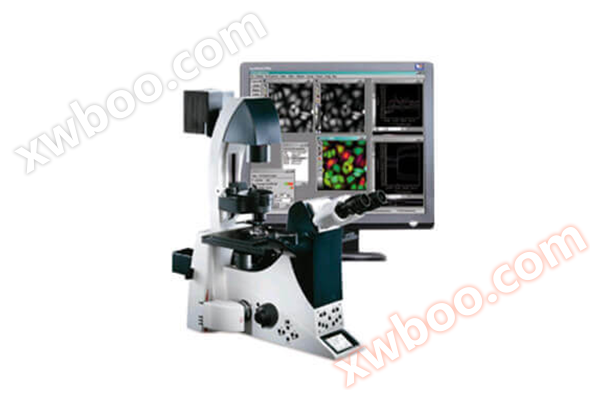
- MetaMorph research grade imaging software
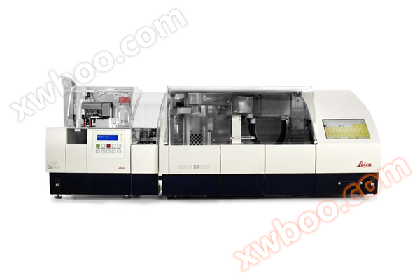
- Dyeing Machine
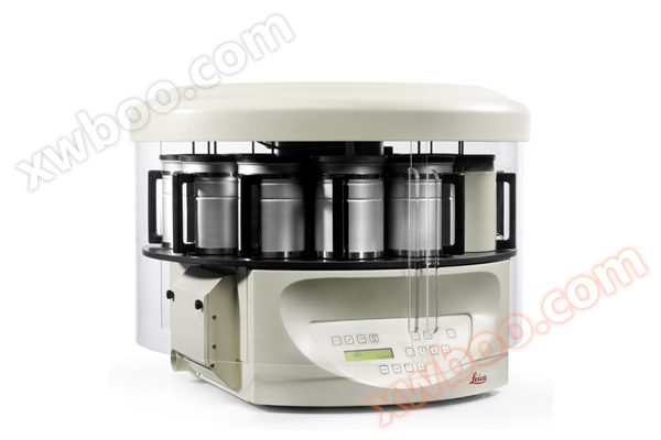
- Modular organization embedding system
- Slide scanner/digital pathology scanner
- Multi functional enzyme-linked immunosorbent assay (ELISA) reader
- Slicer/Oven
- Single function enzyme-linked immunosorbent assay (ELISA) reader
- Upright microscope series
- metallurgical microscope
- Digital Interactive Classroom
- Microscopic digital analysis system
- Number machine
- High connotation imaging analysis system
- Polarizing microscope
- Dehydration machine
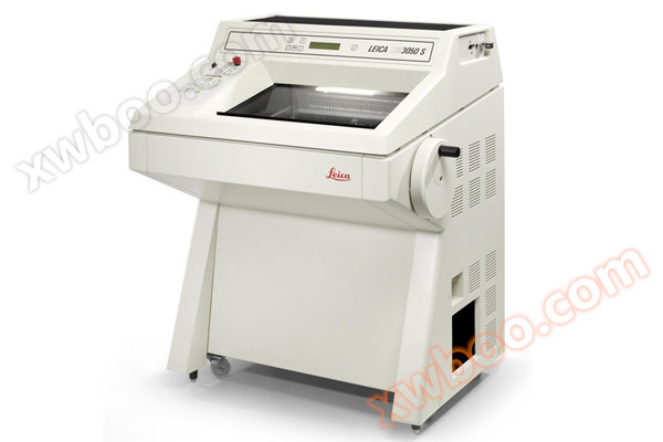
- Slicing machine
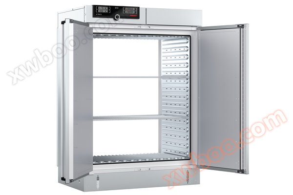
- Memmert oven
Beijing Yuechangxing Technology Co., Ltd

ImageXpress Micro 4 Wide Field High Content Imaging System
NegotiableUpdate on 04/01
- Model
- Nature of the Manufacturer
- Producers
- Product Category
- Place of Origin
Overview
The ImageXpress Micro 4 high content imaging system is designed based on over 30 years of accumulated experience in cell imaging, aiming to improve performance and reliably accelerate your research progress. Combining MetaXpress high content image acquisition and analysis software, The ImageXpress Micro 4 system has become a end-to-end solution that allows you to parse images, understand data, and explore new ideas.
Product Details

ImageXpressMicro4 Wide Field High Content Imaging System
The ImageXpress Micro 4 high content imaging analysis system belongs to the fourth generation imaging technology. Innovative and flexible design, extremely fast operating speed for completing experiments; We also provide you with more options to upgrade the system to confocal imaging level when needed in the future. By jointly using the MetaXpress series of high content image acquisition and analysis software, the ImageXpress Micro 4 system uses intelligent, multidimensional, and high-throughput screening methods to help you discover the next major breakthrough.

main features
Capable of automatically imaging 6-1536 porous plates, including Transwell plates, that meet or do not meet SBS standards for various specimens, specifications, and any bottom wall; And it can preview user made samples such as glass slides, cell chips, and tissue arrays through low magnification mirrors, customize sample specifications, and perform high-throughput automatic imaging and data analysis through high magnification mirrors.
●Scientific grade sCMOS is currently the most advanced detector in microscopy imaging detection systems. SCMOS overcomes the disadvantages of low output rate and high power consumption of CCD, as well as the disadvantages of high noise and dark current of CMOS. It is a scientific grade image detector that integrates high resolution, high frame rate, high quantum efficiency, high sensitivity, high dynamic range, and wide field of view. (Combined with solid-state light engine light source to achieve the best imaging effect).
●Adopting magnetic levitation technology, it has fast speed, high accuracy, no wear, and no thermal fatigue.
●Laser hardware autofocus and software autofocus can be combined and used separately or simultaneously. Flexible application, suitable for various objective lenses and sample plates.
●By using a dedicated phase contrast objective lens, cells without fluorescent labeling can be observed and analyzed. Its phase contrast imaging module can obtain higher contrast unlabeled images, and the light source component has corresponding focusing lenses and phase contrast ring plates to support unlabeled phase contrast imaging, providing high contrast images and enabling cell recognition and tracking in unlabeled conditions.
●PowerCoreTM data parallel analysis function can call local and network idle CPU thread resources, which can improve image analysis speed by several times.
●Various experimental parameters in complex 3D structures can be analyzed through simple mouse operations. Combined with MetaXpress's automatic Z-axis layer scanning, 3D display, and patented Best focus algorithm, porous plates can be used to achieve high-throughput 3D shooting and analysis in a streamlined manner.

application area
It can be used from cells, tissue sections to small model animals. The ImageXpress Micro-4 system obtains the necessary images and quantitative analysis results for the experiment. Capable of quantitative analysis of cell morphology, growth, differentiation, migration, apoptosis, toxicity, metabolic pathways, and signal transduction, suitable for research directions such as cell biology, toxicology, pharmacology, physiology, genetics and embryology, neurobiology, oncology, etc.

Experimental examples
Batch dynamic observation of Transwell experiments.
The principle of Transwell invasion experiment: a membrane with small pores is used to separate different components of the culture medium, and cells are placed in the upper layer of the culture medium. Due to the induction effect of relevant components in the lower layer of the culture medium, cells will migrate from the upper layer of the culture medium to the lower layer of the culture medium. Apply a layer of matrix glue on the membrane to mimic the extracellular matrix, so that cells can digest the matrix and complete the migration process from the upper layer to the lower layer. By detecting the amount of cells in the lower culture medium, the invasion ability of cells can be estimated.
The traditional Transwell experiment uses a microscope for observation, which has low flux, slow speed, and cannot observe the dynamic changes of cells in real time. IXM-4 has excellent laser autofocus and image autofocus capabilities, which can automatically complete Transwell's focusing observation with fast speed and high accuracy, suitable for high-throughput batch observation data. If equipped with environmental control components, it can dynamically observe the migration process of cells for a long time.