- Phone
-
Address
Building 1, International Entrepreneurship Park, No.2 Shangdi Information Road, Haidian District, Beijing
Product Categories
- MetaMorph research grade imaging software
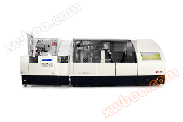
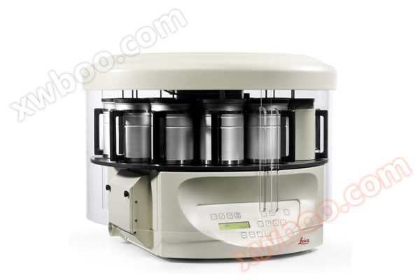
- Dyeing Machine
- Modular organization embedding system
- Slide scanner/digital pathology scanner
- Multi functional enzyme-linked immunosorbent assay (ELISA) reader
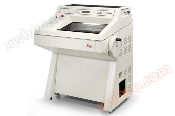
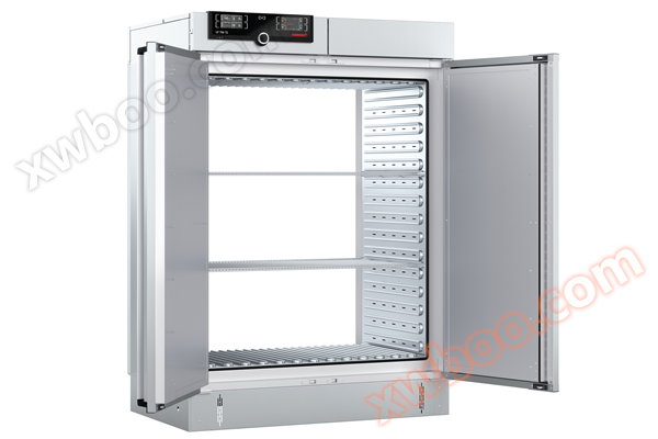
- Slicer/Oven
- Single function enzyme-linked immunosorbent assay (ELISA) reader
- Upright microscope series
- metallurgical microscope
- Digital Interactive Classroom
- Microscopic digital analysis system
- Number machine
- High connotation imaging analysis system
- Polarizing microscope
- Dehydration machine
- Slicing machine
- Memmert oven
Beijing Yuechangxing Technology Co., Ltd

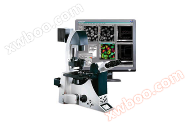
DM6 FS Biological Microscope
NegotiableUpdate on 04/01
- Model
- Nature of the Manufacturer
- Producers
- Product Category
- Place of Origin
Overview
Top level upright microscope for electrophysiological research, intelligent operation, constant color temperature halogen lamp transmission illumination, built-in 750-850mm infrared filter, electrically encoded fluorescence light path, electrically focused, stroke 12mm, optional fixed stage
Product Details

Leica DM6 FS Biological Microscope Suitable for Electrobiology and Neurobiology
If you are seeking a microscope system that supports neuroscience, evolutionary biology research, or in vivo imaging, the Leica DM6 FS fixed stage wide field microscope is your top choice. When you want to stimulate samples in optogenetics or measure samples in electrophysiological experiments such as patch clamp, this wide field microscope shows significant advantages.
Overview of the Advantages of Leica DM6 FS Biological Microscope
During the control process, there is no vibration to ensure that the sample is not disturbed.
When not in use, all current carrying components can be turned off to prevent damage to the sample.
Larger gaps allow for easy access to the sample.
The Leica DM6 FS uses the same bracket as the Leica DM6 B and is an excellent tool for conducting complex and precise experiments. It is also an important component of the Leica EM Cryo CLEM system.

Transform your microscope into an imaging system
Leica Microsystems provides powerful cameras and workflow based software to build integrated imaging systems.
Leica X (LAS X) is a simple and easy-to-use workflow based software that helps you capture images and quickly guides you through operations.
Leica microscope cameras have a wide range of applications, covering various fields from brightfield to fluorescence - even one camera can be used for two applications, such as the Leica DFC7000 T multifunctional camera.

Preparation of samples by laser micro cutting
Accelerate sample preparation by directly obtaining pure and pollution-free initial materials for molecular biological analysis from tissue sections! Laser microdissection (LMD) can separate target cells from other parts of the sample, preserving their complete morphology and helping you accurately observe the area of interest.
Our laser micro cutting system features
We are moving the laser, not the sample. Therefore, our cutting process is very fast and neat.
We use gravity to directly collect the excised tissue in standard consumables, saving time and money.
● Can cut various preparation materials: fresh, frozen, fixed or immunologically labeled samples, live cells, smear preparation materials, bones, plants, wood, dentin, etc.